قال تعالي ( ( وَفِي أَنْفُسِكُمْ أَفَلا تُبْصِرُونَ ) سورة الذاريات ايه ٢١
سوائل الجسم تجري خلال شبكات وبهذة الشبكات محابس تنظم هذا السريان وهذة نعمة من نعم الله تعالي علينا
المصرات عبارة عن عضلات دائرية خاصة تفتح وتغلق أجزاء معينة من الجسم. غالبًا ما يكون عمل العضلة العاصرة هو تنظيم مرور بعض أنواع السوائل ، مثل الصفراء أو البول أو البراز.
العضلة العاصرة هي عضلة دائرية تحافظ عادة على انقباض ممر أو فتحة الجسم الطبيعية والتي ترتاح حسب ما تتطلبه الوظائف الفسيولوجية الطبيعية.
المصرات عبارة عن عضلات تشبه الحلقة تحافظ على انقباض ممر الجسم. هناك العديد من المصرات في جسم الإنسان ، بما في ذلك تلك التي تتحكم في إفراز البول والبراز. توجد في الواقع عضلتان عاصرة في فتحة الشرج - أحدهما داخلي والآخر خارجي.
العضلة العاصرة هي عضلات متخصصة تقع في الجزء العلوي من المريء (العضلة العاصرة للمريء العلوية (UES)) ، والموصل المعدي المريئي (العضلة العاصرة للمريء السفلية (LES)) ، والوصلة العاصرة الإثني عشرية (البواب) ، والموصل اللفائفي (ICJ) ، والشرج (العضلة العاصرة الشرجية) .
تنظيم عمل المصرات
قد يحدث عمل المصرات بشكل لا إرادي من خلال الجهاز العصبي اللاإرادي أو ربما تحت بعض السيطرة الطوعية من خلال الجهاز العصبي الجسدي.
إذا فقدت العضلة العاصرة توتر العضلات أو كان لديها الكثير من التوتر (التشنج) ، يمكن أن تتبع الأعراض و / أو المرض ، مثل احتباس البول وسلس المثانة والبراز.
عضلات الجهاز الهضمي
هناك ستة مصرات مختلفة داخل الجهاز الهضمي. العضلة العاصرة للمريء العلوية يمكن العثور على العضلة العاصرة للمريء العلوية (UES)
Upper Esophageal Sphincter
، والمعروفة أيضًا باسم العضلة العاصرة البلعومية السفلية ، في نهاية البلعوم ، حيث تحمي مدخل المريء.
يعتبر UES مسؤولاً عن منع دخول الهواء إلى المريء عندما نتنفس ومنع استنشاق الطعام إلى مجاري التنفس لدينا.
بسبب موقعه ، يلعب UES أيضًا دورًا في التجشؤ والقيء. يمكن أن يؤدي خلل في UES ، كجزء من مرض الارتجاع المعدي المريئي (GERD) ، إلى عودة الحمض إلى الحلق أو في الشعب الهوائية. العضلة العاصرة للمريء العلوية والارتجاع الحمضي
Lower Esophageal Sphincter
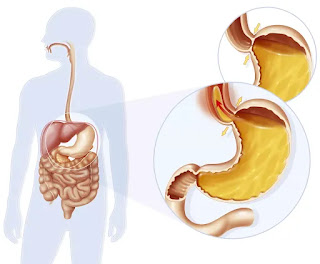
العضلة العاصرة للمريء السفلية تقع المصرة المريئية السفلية (LES) ، والمعروفة أيضًا باسم العضلة العاصرة القلبية ، في الجزء السفلي من المريء حيث تلتقي بالمعدة. وتتمثل وظائفه الأساسية في السماح للطعام بالمرور من المريء إلى المعدة ، والسماح للهواء بالخروج من المعدة عند التجشؤ ، ومنع حمض المعدة من العودة إلى المريء. يعد خلل في LES أحد الأسباب الرئيسية للارتجاع المعدي المريئي
Pyloric Sphincter
العضلة العاصرة البوابية تقع العضلة العاصرة البوابية بين المعدة والاثني عشر ، وهو الجزء الأول من الأمعاء الدقيقة. تفتح العضلة العاصرة البوابية للسماح للطعام المهضوم جزئيًا (الكيموس) بالمرور من المعدة إلى الاثني عشر لمزيد من الهضم وامتصاص العناصر الغذائية في الجسم.
Sphincter of Oddi
مصرة أودي تقع العضلة العاصرة لـ Oddi (SO) حيث تتصل القناة الصفراوية المشتركة وقناة البنكرياس بالاثني عشر. يفتح SO بعد تناول الطعام للسماح للمادة الصفراوية من المرارة ، والإنزيمات من البنكرياس ، بدخول الاثني عشر لتفكيك مكونات الطعام لامتصاصها في الجسم.
العضلة العاصرة لخلل أودي (SOD) ، اضطراب صحي نادر نسبيًا ، يمكن أن يسبب نوبات من الألم في منطقة الصدر.
Illiocecal sphincter
العضلة العاصرة اللفائفية تقع العضلة العاصرة اللفائفية في مكان التقاء الأمعاء الدقيقة والأمعاء الغليظة. لا يُعرف الكثير عن هذه العضلة العاصرة ، بخلاف أنه يُعتقد أنها تطرد الكيموس من نهاية الأمعاء الدقيقة (الدقاق) إلى الأمعاء الغليظة
العضلة العاصرة الشرجية تقع العضلة العاصرة الشرجية في نهاية المستقيم ، وبالتالي في نهاية القناة الهضمية. تنظم العضلة العاصرة الشرجية عملية تفريغ البراز. يحتوي على مكون داخلي وخارجي. تخضع العضلة العاصرة الداخلية للتحكم اللاإرادي (وبالتالي تمنع البراز من التسرب) ، بينما تكون العضلة العاصرة الخارجية في الغالب تحت السيطرة الإرادية ، مما يسمح بحركة الأمعاء. يمكن أن يتسبب خلل في العضلة العاصرة الشرجية في تسرب البراز ، وهي حالة صحية تُعرف باسم سلس البراز.
المصرات الأخرى توجد مصرات أخرى في جميع أنحاء جسمك. العضلة العاصرة الإحليلي تُعرف هذه العضلة العاصرة أيضًا باسم مجرى البول العاصرة ، وتتحكم في احتجاز البول وإفراغه. مثل العضلة العاصرة الشرجية ، تحتوي العضلة العاصرة في مجرى البول على عضلات داخلية وخارجية ، وهي على التوالي تحت السيطرة الإرادية والطوعية. العضلة العاصرة للقزحية يُعرف أيضًا باسم العضلة العاصرة الحدقة أو الحدقة العاصرة. تنظم العضلة العاصرة إغلاق بؤبؤ العين.
لماذا تكون العضلة العاصرة للمريء العلوية فريدة من نوعها تلعب UES دورًا خاصًا في تنظيم مرور الطعام والسائل إلى الحلق ، ولكنها ليست العضلة العاصرة الوحيدة في الجسم. هناك أيضًا العضلة العاصرة الشرجية ، وهي مجموعة العضلات القريبة من فتحة الشرج التي تنظم مرور البراز خارج الجسم. ثم هناك العضلة العاصرة لـ Oddi ، التي تنظم مرور إفرازات الصفراء والبنكرياس إلى الأمعاء الدقيقة. بينما تظهر المصرات في مناطق مختلفة من الجسم ، فإنها تعمل جميعًا على التحكم في تدفق المواد عبر الأعضاء وفتح أجزاء الجسم المختلفة وإغلاقها. تلعب المصرات دورًا مهمًا في الحفاظ على الجسم سليمًا وصحيًا.
وظيفة المصرات او العضلة العاصرة او محابس الأعضاء
باستثناء العضلة العاصرة الشرجية الداخلية ، تعمل المصرات على منع الحركة الخلفية للمحتويات داخل اللمعة.
تمنع العضلة العاصرة الشرجية الداخلية الحركة غير المنضبطة للمحتويات داخل اللمعة عبر فتحة الشرج.
تمنع العضلة العاصرة المريئية السفلية ارتداد حمض المعدة إلى المريء.
العضلات العاصرة الملساء تتكون العضلة العاصرة الملساء من حلقات عضلية تبقى في حالة تقلص مستمر.
آليات "المزلاج" المتخصصة في الخيوط الانقباضية تمكن المصرات من الحفاظ على نغمة الانقباض لفترات طويلة مع الحد الأدنى من استهلاك الطاقة.
يتمثل تأثير حالة الانقباض المقوي في سد اللومن في منطقة تفصل بين جزأين متخصصين. باستثناء العضلة العاصرة الشرجية الداخلية ، تعمل المصرات على منع الحركة الخلفية للمحتويات داخل اللمعة.
تمنع العضلة العاصرة الشرجية الداخلية الحركة غير المنضبطة للمحتويات داخل اللمعة عبر فتحة الشرج.
تمنع العضلة العاصرة المريئية السفلية ارتداد حمض المعدة إلى المريء.
يؤدي عدم الكفاءة إلى التعرض المزمن للغشاء المخاطي للمريء للحمض ، مما قد يؤدي إلى حدوث حرقة في المعدة وتغيرات خلل التنسج قد تصبح سرطانية.
تمنع العضلة العاصرة المعوية ، والتي تسمى أحيانًا العضلة العاصرة البوابية ، الارتجاع المفرط لمحتويات الاثني عشر إلى المعدة.
يمكن أن يؤدي عدم كفاءة هذه العضلة العاصرة إلى ارتداد الأحماض الصفراوية والإنزيمات المحللة للبروتين من الاثني عشر. الأحماض الصفراوية والإنزيمات المحللة للبروتين تضر الحاجز الواقي في الغشاء المخاطي في المعدة. يمكن أن يؤدي التعرض لفترات طويلة إلى التهاب المعدة وتقرحها.
تمنع العضلة العاصرة اللفائفي القولونية ارتداد محتويات القولون إلى الدقاق.
يمكن أن يسمح عدم الكفاءة بدخول البكتيريا إلى الدقاق من القولون وقد يؤدي إلى فرط نمو البكتيريا.
عادة ما تكون أعداد البكتيريا منخفضة في الأمعاء الدقيقة. (لماذا)
معظم البكتريا تعيش في وسط متعادل والاس الهيدروجيني بالامعاء الدقيقه قلوي
اعجاز
وظيفة الامعاء الدقيقة
وقلويه الامعاء الدقيقة مناعة طبيعية
A sphincter is a circular muscle that normally maintains constriction of a natural body passage or orifice and which relaxes as required by normal physiological functioning. Sphincters are found in many animals.
The action of sphincters may happen involuntarily through the autonomic nervous system or maybe under some voluntary control through the somatic nervous system.
If a sphincter loses muscle tone or has too much tone (spasticity), symptoms and/or illness can follow, such as urinary retention and bladder and fecal
incontinence
Sphincters are special, circular muscles that open and close certain body parts. Most often, the action of a sphincter is to regulate the passage of some type of fluid, such as bile, urine, or fecal matter.
Sphincter
Sphincters are specialized muscles that are located at the upper esophagus (upper esophageal sphincter (UES)), gastroesophageal junction (lower esophageal sphincter (LES)), antroduodenal junction (pylorus), ileocecal junction (ICJ), and the anus (anal sphincter).
From: Reference Module in Biomedical Sciences, 2014
Smooth Muscle Sphincters
Smooth muscle sphincters consist of rings of muscle that remain in a continuous state of contraction. Specialized “latch” mechanisms in the contractile filaments enable sphincters to maintain contractile tone for extended periods with minimal expenditure of energy. The effect of the tonic contractile state is to occlude the lumen in a region that separates two specialized compartments. With the exception of the internal anal sphincter, sphincters function to prevent the backward movement of intraluminal contents. The internal anal sphincter prevents uncontrolled movement of intraluminal contents through the anus.
The lower esophageal sphincter prevents reflux of gastric acid into the esophagus. Incompetence results in chronic exposure of the esophageal mucosa to acid, which can lead to heartburn and dysplastic changes that may become cancerous. The gastroduodenal sphincter, which is sometimes called the pyloric sphincter, prevents excessive reflux of duodenal contents into the stomach. Incompetence of this sphincter can result in the reflux of bile acids and proteolytic enzymes from the duodenum. Bile acids and proteolytic enzymes are damaging to the protective barrier in the gastric mucosa; prolonged exposure can lead to gastritis and ulceration.
The ileocolonic sphincter prevents reflux of colonic contents into the ileum. Incompetence can allow entry of bacteria into the ileum from the colon and may result in bacterial overgrowth. Bacterial counts are normally low in the small intestine.
...
Digestive System Sphincters
There are six different sphincters within the digestive system.
Upper Esophageal Sphincter
The upper esophageal sphincter (UES), also known as the inferior pharyngeal sphincter, can be found at the end of the pharynx, where it protects the entrance to the esophagus. The UES is responsible for preventing air from getting into the esophagus when we breathe and to prevent food from being
aspirated into our respiratory tracts.
Because of its location, the UES also plays a role in burping and vomiting. Malfunctioning of the UES, as part of gastroesophageal reflux disease, (GERD) can cause acid to back up into the throat or into the airways.
Lower Esophageal Sphincter
The lower esophageal sphincter (LES), also known as the cardiac sphincter, is located at the bottom of the esophagus where it meets up with the stomach.
Its primary functions are to allow food to pass from the esophagus into the stomach, to allow air to escape from the stomach when burping, and to prevent stomach acid from washing back up into the esophagus. A malfunction of the LES is one of the primary causes of GERD.2
Pyloric Sphincter
The pyloric sphincter is located between the stomach and the duodenum, which is the first part of the small intestine. The pyloric sphincter opens to allow partially digested food (chyme) to pass from the stomach into the
duodenum for further digestion and absorption of nutrients into the body
Sphincter of Oddi
Sphincter of Oddi (SO) is located where the common bile duct and the pancreatic duct connect to the duodenum. The SO opens after we have eaten so as to allow bile from the gallbladder, and enzymes from the pancreas, to enter the duodenum so as to break down food components for absorption into the body.
Sphincter of Oddi dysfunction (SOD), a relatively rare health disorder, which can cause episodes of pain in the chest area.3
Ileocecal Sphincter
The ileocecal sphincter is located where the small intestine and the large intestine meet. There is not much known about this sphincter, other than that it is thought to expel chyme from the end of the small intestine, (the ileum) into the large intestine.
Anal Sphincter
The anal sphincter is located at the end of the rectum, and therefore at the end of the digestive tract. The anal sphincter regulates the process of the evacuation of stool. It has both an inner and outer component.
The inner sphincter is under involuntary control (and therefore prevents stool from leaking out), while the outer sphincter is predominantly under voluntary control, thus allowing for a bowel movement. A malfunction of the anal sphincter can cause stool leakage, a health condition known as fecal incontinence.4
Other Sphincters
There are other sphincters that you have throughout your body.
Urethral Sphincter
Also known as the sphincter urethrae, this sphincter controls the holding and emptying of the urine. Like the anal sphincter, the urethral sphincter has both inner and outer muscles, which are respectively primarily under involuntary and voluntary control.
Iris Sphincter
Also known as the pupillary sphincter or sphincter pupillae. This sphincter regulates the closing of the pupil in the eye
How the Upper Esophageal Sphincter Works
During swallowing, the upper esophageal sphincter opens to allow food and liquids to pass into the esophagus.1 It can also reduce the backflow of food and liquids from the esophagus into the pharynx.
In addition to eating, we use this part of the esophagus while simply breathing. It also comes into play during unpleasant bodily functions, such as burping or throwing up, that serve to expel gas or harmful materials from the body.

ليست هناك تعليقات:
إرسال تعليق